Blindness Simulator
When someone you care about experiences vision loss, it can be hard to understand exactly what they’re going through. Vision changes aren’t always obvious—and they don’t look the same for everyone. Some people may lose central vision, others may have trouble seeing at night, and some may see the world as blurry or foggy. This guide is designed to help families, friends, and caregivers better understand the different types of vision loss. At ACBVI, we believe that knowledge builds empathy—and we’re here to support both individuals with vision loss and the people who walk beside them.
Hover or focus on each card to see how different vision conditions affect what you see.

Central Vision Loss
A noticeable blur or complete loss of vision in the center of your visual field. This makes reading, recognizing faces, or focusing on details directly ahead difficult—even if side vision remains intact.
Peripheral Vision Loss
A narrowing of your visual field where side (peripheral) vision fades or disappears, often described as looking through a tunnel. Central vision may remain sharp, but navigating crowded spaces or detecting movement from the sides becomes challenging.
Media Opacity
A foggy or cloudy view caused by obstructions in the eye’s clear structures—such as the lens (cataracts), cornea (scars), or the vitreous (gel inside the eye). These changes scatter light and blur vision in ways that glasses or contacts can’t fix. People may also experience multiple overlapping types of vision loss.
Early Stage Macular Degeneration
Subtle central vision changes such as wavy lines, faded colors, or small blurry patches. These symptoms may be mild but are early signs of damage to the macula, the part of the retina responsible for sharp detail.
Late Stage Macular Degeneration
Advanced damage to the macula leads to large, dark, or blank areas at the center of vision. It becomes extremely difficult or impossible to read, drive, or recognize faces, although peripheral vision may still function.
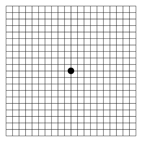
Amsler Grid
A simple, square grid with a central dot used to check for vision distortions or blind spots. If lines appear wavy or parts of the grid are missing, it may indicate central vision problems like macular degeneration.
Incomplete Scotoma
A partially blocked area in the visual field—like a blurry smudge or faded spot—where sight is reduced but not totally gone.
Ring Scotoma
A ring-shaped blind zone surrounding the central field of vision. People may see clearly in the very center and far periphery but miss everything in between—creating a “doughnut” effect of vision loss.
Absolute Scotoma
A completely blank or black area in the field of vision. No light or shapes are perceived in this zone, and it often results from localized retinal or brain damage.
Retinitis Pigmentosa (RP)
A group of inherited eye disorders that gradually damage the retina. Night vision is usually affected first, followed by a slow loss of peripheral vision, eventually resulting in tunnel vision.
RP/Glaucoma
A compounded vision loss where Retinitis Pigmentosa and glaucoma both affect the eye. The result is widespread vision dimming, blind spots, and pressure-related damage—impacting both clarity and field.
Homonymous Visual Field Loss
Loss of the same side of the visual field in both eyes—typically from a brain injury or stroke. For example, you might be unable to see anything to your right even though both eyes are open and healthy.
Homonymous Hemianopsia
Complete blindness in the left or right half of your vision in both eyes. It’s not an eye problem but usually the result of brain damage on one side affecting how vision is processed.
Left-Side Loss with Macular Involvement
Loss of vision on the left side of your visual field that also includes damage to the central (macular) area, impairing both detail and orientation.
Left-Side Loss with Macular Sparing
A blind area on the left side of vision, but the central field (macula) remains unaffected—preserving the ability to see straight ahead while side vision is reduced.
Diabetic Retinopathy
A diabetes-related condition where blood vessels in the retina leak or swell, causing blurred vision, floaters, and even permanent damage. Vision may fluctuate or decline unpredictably.
Eye Hemorrhage
Bleeding within the eye—often from trauma or medical conditions like diabetes—leads to sudden vision disturbances such as dark blotches, veils, or flashes that block parts of your view.
Need more information?
If you have any questions or need additional information, we encourage you to reach out to us at ACBVI. Our team is dedicated to assisting you and providing the resources and support you require. Don’t hesitate to get in touch—we’re here to help. Contact us by phone at 602-273-7411 or via email at info@acbvi.org. To stay up to date with the latest news, subscribe to our newsletter.